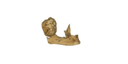
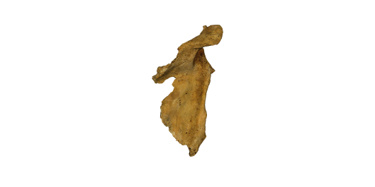
On ethics, fair use and 3D printing

Digitised Diseases is an online digital resource of pathological type specimens made up of more than 1600 3D models of human remains. Given the sensitive nature of the content and the type of collections that we have accessed as part of this project we have an obligation to remind users that whilst they are free to access content for non-commercial educational purposes, any misuse of material (e.g. creation of artwork/ installations or printing of 3D models) from this resource will be taken very seriously. The Royal College of Surgeons in particular is subject to a license from the Human Tissue Authority (HTA) and is obliged to inform the HTA of any misuse of images involving their modern human remains. We would therefore ask that users are respectful in their use of this content, mindful that these are bones of real people and that users approach the resource with the same reverence that they would afford actual human remains irrespective of age.

This statement will eventually appear on the Digitised Diseases website.

Project Launch Day

After a lovely write-up in the Guardian yesterday we’ve had lots of interest in the project.

The website will be going live at 1700 GMT just ahead of the launch at the Royal College of Surgeons this evening (invite only, sorry!).

Digitised Diseases will be available at digitiseddiseases.org 

1 day to go until the Launch


B046000


Left ulna


The shaft is primarily absent, with only a small portion of the proximal end present, due to amputation.  The distal extent of the bone is diminished and has an irregular pitted surface. There are three anterior projections of bone, one below the coronoid process and two distal to the radial notch on the lateral border of the shaft.


When compared to the right side the shoulder, arm and forearm shows considerable atrophy, most likely from disuse.


The level of amputation on the ulna is at that of the radius exactly.  The individual may have lost their arm in an accident, rather than it being surgically removed.


Disease Classification:


Traumatic-surgical-amputation

B015700


First lumbar vertebra


There are irregular indentations on the inferior and superior surfaces of the body in the centre of the posterior half.  The lesions retain the original cortical bone for most of the floor.  The superior indentation measures at its greatest 18.3mm mediolateral x 9.8mm anteroposterior x 4mm deep.  There is a small nodule of new compact bone on the left lateral border of the lesion, minimal trabecular exposure and a small area of irregular coarse pitting centrally in the base. The inferior indentation is crescent shaped and has more trabecular exposure than the superior lesion and measures at its greatest 16.9mm mediolateral x 5mm anteroposterior x 3.3mm deep.


Disease Classification:


Degenerative joint disease-Schmorl’s node

B019900


Right femur


There is a well healed transverse fracture at midshaft with complete posteromedial displacement of the distal shaft. The medial half of the proximal end of the shaft has remodelled compact bone. A distal extension of remodelled compact bone that surrounds the shafts creates a bridge between the fractured halves, except on the distolateral half of the posterior surface where the space between the two halves is filled with irregular spicules of new compact bone, having a minimal amount of pitting.  There is slight pitting on the anterior surface superior to the fracture.


Co-existing Pathology:


There is slight osteophytosis of the femoral head.  The head also has surface contour changes superior to the fovea, including a depression measuring 5mm mediolateral x 9.6mm anteroposterior.   Osteophytic extensions of the articular surface of the head bridge onto the neck on both superoanterior and superolateral aspects.


The distal articulation has extensive osteophytosis with the patellar surface having contour change, surface erosion, new compact bone formation and pitting.  The lateral condyle has a large irregular osteophyte interrupting the medial margin and the articular surface has new compact bone and surface pitting.  The medial distal portion of the medial condyle has extensive eburnation and grooving, with a nodule of new compact bone anterior to this area and a zone of pitting and contour changes within the medial half of the eburnated area.


The osteoarthritic changes of the distal femur and lack of atrophy of the bone and muscle attachments suggests that the individual had been using their leg for some time after fracturing the shaft.


Disease Classification:


Trauma-accidental-fractures


Degenerative joint disease-osteoarthritis


Degenerative joint disease-osteophytosis

B035700


Right femur


Distal to midshaft along the lateral side of the linea aspera at the attachment for quadriceps and biceps femoris is a large exostosis of bone measuring 45mm proximodistal x 19.5mm anteroposterior x 8.2mm mediolateral at its greatest. There is continuity from the cortical bone to the exostosis.


The x-ray confirms that this is an osteochondroma.


Disease Classification:


Neoplastic-benign-osteochondroma

B039700


Left radius


There is a well-healed fracture at midshaft. The distal portion is displaced anteriorly and proximally, and is angled medially.  There is little evidence of a callus remaining.


Disease Classification:


Traumatic-accidental-fractures


Count down to the launch 4 days…

B131400

Left femur

The distal third of shaft is greatly expanded mediolaterally and is highly porotic (being more extensive than normal development).  The whole of the shaft is bowed anteroposteriorly and the distal end is medially rotated.  There is a large area of new woven bone on the proximal third of the shaft.

Disease Classification:

Metabolic-deficiency-rickets

M004000

Eighth thoracic to the fourth lumbar vertebrae

The bodies and apophyseal joints of the eighth thoracic to fourth lumbar vertebrae are fused. Bone destruction in the tenth thoracic to first lumbar bodies has resulted in severe anterior kyphosis (c 140 degrees), corresponding to Pott’s disease of the spine. A cloaca drains from the anterior body of the fourth lumbar vertebra.

Co-existing Pathology:

There is mild marginal osteophytosis evident on the superior body of the eighth thoracic vertebra, possibly a consequence of the severe changes in the spine caused by Pott’s disease.

Disease Classification:

Infective-specific-bacterial-tuberculosis

Degenerative joint disease-osteophytosis

M024500

Mandible

There is a sub-oval growth of compact bone (40mm mediolateral, 46.2mm anteroposterior, 44.2mm superoinferior) with a flattened superior surface attached to the left mandibular condyle. The surface of the growth is formed of smooth and regular cortical bone with two large folds on the medial aspect and a build-up of spicular bone on the lateral surface. In contrast, the superior surface of the deposit is slightly concave and consists of irregular, uneven lamellar bone which is spongy in appearance. This may reflect the outline of the bone and soft tissue architecture at the point of contact with the temporal area.

Considering the age of this individual, there is relatively little wear on the occlusal surfaces of the teeth, and the left mandibular molars are slightly more worn than the right. It is possible that the pathological lesion reduced masticatory function throughout the jaw. The radiograph reveals a disorganised trabecular internal structure with an irregular radiodense area along the superior border, which was presumably in contact with the temporal bone.

The lesion is large, continuous with the medullary cavity, arises from a physeal area and contains trabecular bone surrounded in the main by cortical bone. These are all characteristic of osteochondromas which occasionally occur in the mandible. This lesion would have caused progressive facial asymmetry.

Disease Classification:

Neoblastic-benign-osteochondroma

M027000

Cranium

There is a flattened spherical mass of bone, 34mm in diameter and extending 18mm above the normal bone surface. This is attached to the right parietal notch, and overlaps the temporal bone just above the mastoid process. The lesion is lobulated with a smooth surface of dense lamellar bone and occasional porosity. The majority of the base of the growth lies above the normal bone surface. No bone changes are present on the endocranial surface.

Disease Classification:

Neoplastic-benign-osteoma

Coming Soon…

B120200

Mandible

There is a large lobulated mass of compact bone on the right ascending ramus.  The mass measures c. 56.4mm superoinferior x 42.1mm anteroposterior x 30mm mediolateral.  The medial surface of the assenting ramis is heavily pitted.

Co-existing Pathology:

Teeth lost ante-mortem: LL3,5,6: LR4,5,6; Teeth lost post-mortem: LL1,2,3,4,7,8: LR 1,2,3,4,,7,8.  Lower left 7 and LR7 have severe mesial tilt indicating LL6 and LR6 had been lost when individual aged 12 -18 years.

Disease Classification:

Neoplastic-benign-osteoma

Miscellaneous-dental-ante-mortem tooth loss
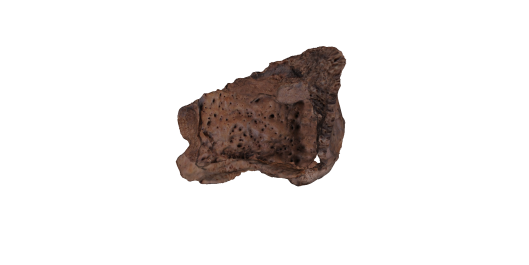
B009300

Right maxilla

This is a posterior fragment of the right maxilla. There is a layer of new compact bone, which is nodular, on the visible surfaces with extensive pitting (the pits being large in size).

Disease Classification:

Infective-non-specific-maxillary sinusitis

b010300

Frontal and maxillae

Orbits- There is a diffuse convoluted mixture of bone destruction, new compact bone formation and expansion of the diploic channels on the right orbital roof and on an area of the greater wing of the sphenoid superiorly. The left orbit is covered in remodelled smooth new compact bone with areas of bone erosion and expansion of the diploic channels giving a convoluted appearance.

Maxillae- There is new compact bone on the nasal floor that has a vascular pattern and extensive porosity near the right infra-orbital foramen.  There is extensive pitting of the anterior quarter of the palate.

Zygoma- There is slight pitting and bone erosion bilaterally.

Sphenoid-There is mild pitting to the external surface of the right greater wing.

Frontal- There is a large area of bone destruction on the endocranial surface of the frontal. This lesion is located centrally on the superior and anterior surface. There is expansion of the diploic channels transversely and perpendicular expansion of trabeculae through the endocortical surface centrally and superiorly within the lesion, with convoluted areas of cortical bone anteriorly and laterally.

Disease Classification:

Metabolic-deficiency-anaemia

Miscellaneous-skeletal-cribra orbitalia

Miscellaneous-skeletal-endocranial lesions

B031000

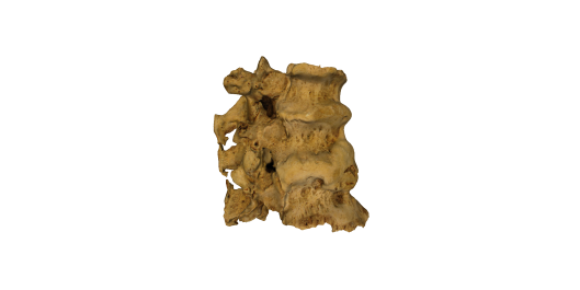
Tenth to twelfth thoracic vertebrae and a partial left tenth rib

The tenth to twelfth thoracic vertebrae are fused though a flowing ossification of the anterior longitudinal ligament, which becomes a more irregular ossification on the right side on the twelfth thoracic vertebra and on the left side of the tenth to twelfth thoracic vertebrae.  This flowing ossification also extends to and fuses the left tenth rib to the vertebral body of tenth thoracic vertebra, which is broken post-mortem.  An extension of bone from twelfth thoracic to first lumbar vertebrae creates a pseudo-articulation.  The intervertebral disc spaced is preserved.  There is a flowing ossification of ligaments from below the transverse process of eleventh thoracic, over the joint, to just superior to the right inferior facet of the twelfth thoracic vertebra.

The vertebrae would have had continuous fusion from the second to tenth thoracic vertebrae.  However, it is broken post-mortem into three separate pieces.

Co-existing Pathology:

Osteophytosis is visible on the exposed vertebral body margins, which in many places can be observed to be overlapped by the flowing ossification of the anterior longitudinal ligament.

Other pathological changes include variations in the surface contour of the laminae of most of the vertebrae; all costal facets have osteophytic lipping and joint surface contour changes;  partial ossification of the supraspinous ligament from tenth to twelfth thoracic vertebrae, with fusion between tenth and eleventh thoracics; surface pitting on the exposed superior surface of fifth thoracic vertebra; and a nodule and an irregular plaque of new compact bone anterior to the posterior margin of the inferior surface of the body of ninth thoracic vertebra.

When are articulated, the vertebral column shows kyphosis and left scoliosis—with an apex of curvature at the fourth and fifth thoracic vertebrae—and abnormal lordosis in the lower lumbar.

Disease Classification:

Miscellaneous-skeletal-diffuse idiopathic skeletal hyperostosis

Miscellaneous-skeletal-enthesophytes

Degenerative joint disease-osteoarthritis

Degenerative joint disease-osteophytosis

B110300

Scapula

The majority of this bone is covered in multiple fine osteolytic lesions and a layer of coarse woven bone, sparing only areas of the blade and glenoid fossa.   Taphonomic damage obscures the extent of the new bone formation; it is likely to have been more extensive.

The lesions presented here are the result of metastatic carcinoma of the prostate.  However, there is a mixture of pathological lesions distributed throughout the skeleton that are consistent with the individual having co-existing metastatic carcinoma and leprosy.  Elements of the lower limb that have leprous lesions or a mixture of lesions from both diseases have been omitted from this project to prevent confusion.  Metastatic carcinoma of the prostate is characterised by osteosclerotic lesions and deposition of periosteal new bone (Ortner et al 1991).

References Cited:

Ortner DJ, Manchester K, Lee F.  1991. Metastatic carcinoma in a leper skeleton from a medieval cemetery in Chichester, England.  International Journal of Osteoarchaeology 1: 91-98.

Disease Classification:

Neoplastic-malignant-metastatic-metastatic carcinoma
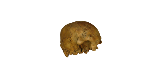
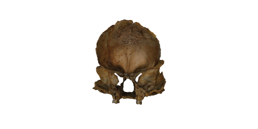
B115800

Cranium

Multiple ectocranial osteoblastic lesions are located across the cranium, focused mainly on the frontal and occipital.  The majority of these bony protrusions are smooth and contiguous with the original/normal cortical surface. However, two located on the occipital are partially lobulated in appearance, with defined inferior margins. The frontal has at least five lesions: one on the right zygomatic process, one on the right temporal line, one adjacent to the left temporal line, one inferior to glabella on the left side, and one adjacent to the coronal suture on the right side.  Here, the largest protrusion is on the right supraorbital margin/zygomatic process and measures c. 19.2mm mediolateral x 24.4mm superoinferior.  Two protrusions are visible on the right and one on the left parietal adjacent to the lambdoid suture.  The occipital has at least seven large protrusions, five occurring centrally along sutra mendosa, the largest being c.25mm in diameter, and two adjacent to the lambdoidal suture.  In this same area on the left side is a cluster of very small irregular nodules of bone.

The diagnosis of this pathology remains uncertain.  Ortner (203, 521-524) present a similar case, which is diagnosed as fibro-osseous tumours.  These tumours are benign and slow growing.

Co-existing Pathology:

Bilaterally are central areas of joint surface destruction on the articular tubercles of the tempomandibular joint (the left side is partially obscured by taphonomic damage).

References Cited:

Ortner DJ. 2003. Identification of pathological conditions in human skeletal remains. London: Academic Press.

Disease Classification:

Developmental-dysplastic-fibro-osseous tumours

Coming soon to a computer near you…

B006400

Right humerus


The majority of the distal half of the bone is covered by new compact and woven bone.  Much of the central anterior half of this bone fragment and the anterior joint surface is unaffected.


Anterior surface- There is a large oval destructive lesion with sharp margins on the lateral border of the distal quarter of the shaft that penetrates an area of porous new woven bone. This woven bone overlies compact bone and the destructive lesions extend deep into the cortical surface leaving some trabeculae exposed.  The remaining distal end is a mix of bone destruction and irregular porous new compact bone deposition with areas of overlaying new woven bone. There are nodules of new compact bone at the common extensor tendon attachment. There is a septal aperture present allowing for an uninterrupted continuation of the new compact bone from the anterior to the posterior surface.


Posterior surface- There is a large amount of new compact bone mixed with porous woven bone covering the entire surface, causing the shaft to be considerably expanded posteriorly. There are multiple gummatous lesions (destructive lesions) in varying stages of activity and healing covering the inferior third of the bone.  Many of these lesions penetrate into the new bone, with some continuing into the cortical bone surface.  The majority of these lesions retain a sharp and irregular margin.  The olecranon fossa is bordered by striated new compact bone.  Approximately half of the lateral epicondyle has been destroyed posteriorly. There is a large spherical destructive lesion lateral to the trochlea that penetrates into the trabeculae, which has sharp margins and a smooth floor.  There is irregular joint surface destruction on the lateral half of the surface of the trochlea.


Radiographs reveal multiple ovoid translucencies with indeterminate margins that are the result of cortical destruction, rather than being internal voids.


Disease Classification:


Infective-specific-bacterial-treponemal

B006500

Left tibia

There is a mixture of compact bone and woven bone covering the majority of the distal third of the shaft, thickening the distal end, especially the anterior surface.

Anterior, medial and lateral surfaces- There is a large focal area of bone deposition in the superior half of the affected area of the shaft. This consists of a mixture of new compact bone and striated porous woven bone.  There are approximately eight small ovoid lytic lesions (gummatous lesions) that have sharp and well defined margins, which penetrate into the original cortical bone surface.  There is a larger lytic lesion (gummatous lesion) laterally that has less defined, irregular margins and floor.  Inferior to this large focal area the bone surface is covered in an irregular mixture of striated and smooth new compact bone, with a large portion on the medial surface having new woven bone overlaying.

Posterior surface- There is layer of striated new compact bone on the distal quarter of the shaft.

Radiographs reveal multiple ovoid translucencies with indeterminate margins that are the result of cortical destruction, rather than being internal voids.

Disease Classification:

Infective-specific-bacterial-treponemal

B006800

Cranium

There is a large active lytic lesion on the right side of the frontal superior to the super-orbital ridge, which penetrates from the outer table to the diploë (the inner table is still intact). It is a square circumvallate cavitation measuring 19.7mm at its largest diameter.  The superior margin is still sharp and the lesion is surrounded by pitting.  The remaining margins are rounded with a rim of new compact bone.  The floor of the lesion shows a mixture of bone destruction and nodular new compact bone formation. Inferior and lateral to this lesion is another that is in a more advanced state of healing. It measures 11.8mm at its maximum diameter and consist of rounded margins of new compact bone and large nodules of new compact bone filling the floor of the lesion.  Superior to glabella are two discrete depressions with radial scarring, with the inferior lesion having pitting. On the posterior surface of the right parietal near the sagittal and lambdoid sutures, there is a large depression with healed new compact bone and radial scarring.  Similarly, there is a healed radial lesion on the occipital above the nuchal crest.

Radiographs reveal multiple ovoid translucencies with indeterminate margins that are the result of cortical destruction, rather than being internal voids.

Co-existent Pathology:

There is bilateral pitting on the superior surface of the orbit.  The surfaces of the posterior parietals, the parietals along the sagittal suture, the right temporal, the right greater wing of the sphenoid, and the occipital are densely pitted.

Disease Classification:

Infective-specific-bacterial-treponemal

Miscellaneous-skeletal-cribra orbitalia

B007100

Sternum (manubrium and sternal body are fused)

There is a thick layer of irregular and porous new compact bone on all non-articular surfaces of the sternal body and manubrium.  The anterior surface of the manubrium has a thick layer of irregular and porous new compact bone and at least three zones of bone destruction.  The most superior and lateral lesion on the left is spherical and has sharp irregular margins and a smooth base.  The other two lesions are irregular in shape, penetrate the cortical surface and are filled with irregular new bone formation.  The right side of the manubrium is covered primarily in layers of thickly striated new compact bone. The anterior surface of the body is primarily covered in a layer of smooth new bone with areas of porosity and pitting (especially in the superior third).  The entire posterior surface of the manubrium and body is covered in multiple layers of irregular and highly porous new compact bone.   There is destruction to much of the left and right superior clavicular articular surfaces.  A circular destructive lesion on the right clavicular articular surface has sharp margins and a large underlying spherical cavity with smooth sides.

Radiographs reveal multiple ovoid translucencies with indeterminate margins that are the result of cortical destruction, rather than being internal voids.


Disease Classification:

Infective-specific-bacterial-treponemal


Coming soon to your computer…

B0080

Left femur

On the distal third of the shaft are two large cloacae that drain from the medullary cavity to both the anterior and posterior surfaces. The posterior cloaca measures 46mm proximodistal x 26mm mediolateral and has smooth margins.  Within the distal half of the cloaca is a large irregular necrotic bone fragment (sequestrum) adhering to the margin, which measures 23mm proximodistal x26mm mediolateral.    There is also small sequestrum adhering to the superior margin.  The cloaca on the anterior surface measures 15mm proximodistal x 13mm mediolateral and has smooth rounded margins.  The cloacae are surrounded by a large involucrum that extends the distal two thirds of the shaft. The involucrum consists of a mixture of smooth and irregular compact bone deposited on the original cortical surface and highly disorganised spicules of new compact bone on the posterior surface.  On the lateral side of the posterior surface is a large disorganised extension of bone that has two perforating channels for nervous and vascular supply.  The majority of the new compact bone on the anterior surface is smooth. However, there are areas of irregular deposits of nodular compact bone.  There is a faint trace of a fracture line running superolaterally to inferomedially (confirmed by radiographs).  This is osteomyelitis secondary to a compact fracture.

Co-existing Pathology:

There is extensive osteophyte formation creating a bony ring surrounding the distal joint surface.

Disease Classification:

Infective-non-specific-osteomyelitis

Traumatic-accidental-fractures

Degenerative joint disease-osteophytosis

b000300

Right os coxae

There is spiculated new compact bone formation on the auricular surface, which has an irregular joint surface contour.  Superoposterior to this, there is an elliptical area of bone destruction, which has smooth margins and that interrupts a portion of the posterior iliac crest.  There is a channel of reactive new compact bone and destructive porosity between the auricular surface to the inferior margin of the destructive lesion. These changes are evidence of tuberculosis of the sacroiliac joint. On the gluteal surface, there is a large, but localised, area of pitting and spiculated new compact bone.

Disease Classification:

Infective-specific-bacterial-tuberculosis

B001000

Fourth and Fifth lumbar vertebrae

There is a destructive lesion on the mid-section of the anterior surface of the body with spiculated new compact bone at its base.  There is ossification of the anterior longitudinal ligament on the left side (less extensive than B0011) joining that of the fourth lumbar vertebra.  This bony bridge is not fused together, but is a pseudo joint allowing for some movement.  On the right side of the body there is a mixture of new woven bone and ossified ligament.  Along with the changes in the fourth lumbar vertebra, this is possible evidence of a psoas abscess associated with tuberculosis.

Disease Classification:

Infective-specific-bacterial-tuberculosis

Pathology of the “Week”: Neoplasm

This slideshow requires JavaScript.

This cranium is from an individual from Post-Medieval Wolverhampton who has multiple benign fibro-osseous tumours.

Cranium

Multiple ectocranial osteoblastic lesions are located across the cranium, focused mainly on the frontal and occipital.  The majority of these bony protrusions are smooth and contiguous with the original/normal cortical surface. However, two located on the occipital are partially lobulated in appearance, with defined inferior margins. The frontal has at least five lesions: one on the right zygomatic process, one on the right temporal line, one adjacent to the left temporal line, one inferior to glabella on the left side, and one adjacent to the coronal suture on the right side.  Here, the largest protrusion is on the right supraorbital margin/zygomatic process and measures c. 19.2mm mediolateral x 24.4mm superoinferior.  Two protrusions are visible on the right and one on the left parietal adjacent to the lambdoid suture.  The occipital has at least seven large protrusions, five occurring centrally along sutra mendosa, the largest being c.25mm in diameter, and two adjacent to the lambdoidal suture.  In this same area on the left side is a cluster of very small irregular nodules of bone.

The diagnosis of this pathology remains uncertain.  Ortner (203, 521-524) present a similar case, which is diagnosed as fibro-osseous tumours.  These tumours are benign and slow growing.

Co-existing Pathology:

Bilaterally are central areas of joint surface destruction on the articular tubercles of the tempomandibular joint (the left side is partially obscured by taphonomic damage).

References Cited:

Ortner DJ. 2003. Identification of pathological conditions in human skeletal remains. London: Academic Press.

Disease Classification:

Developmental-dysplastic-fibro-osseous tumours

Neoplastic-benign-fibro-osseous tumours

Miscellaneous-temporomandibular joint disease

Pathology of the Week: A Review of Paget’s Disease

This slideshow requires JavaScript.

These specimens all show pathological changes associated with Paget’s Disease.

B0146: B82 EQ 6/7

Partial frontal

There is marked thinning of the inner and outer cortical layers, with extensive surface porosity on all surfaces of the bone fragment.  There is considerable thickening of the cranial vault by endosteal and subperiosteal new bone formation that is highly irregular, disorganised, and fills the diploic space.  The grooves for the meningeal vessels are particularly deep.   At its thickest, the fragment measures 23mm endo- to ectocranially.


B0148: Hull Magistrates Court 1965

Left femur

The shaft and the greater and lesser trochanters are considerably thickened by a deposition of highly disorganised new bone, especially on the medial and lateral surfaces, with substantial narrowing of the medullary cavity.  The external surface of the bone is highly disorganised with pitting and porosity.  There are no changes to the surface of the femoral neck or head, with the trabeculae of the head remaining normal.


B0150: Lincoln SUS ’96 116

Right os coxae

The entire bone is considerably thickened by a deposition of highly porous and pitted disorganised new compact bone mixed with irregular striations of compact bone on all non-articular surfaces.  Two small patches of irregular woven bone are located on the lateral surface near the inferior gluteal line.  The superior margin of the acetabulum has been extended by osteophytic growth. Within the acetabulum are multiple destructive lesions that have irregular sharp margins, many of which perforate into the trabeculae. On the superolateral surface in the acetabulum is a large area of eburnation.


B0733, B0734, B0738: Hereford Cathedral (HE93A) 3051

Left tibia

There is considerable thickening of the proximal half of the shaft with deposition of highly disorganised subperiosteal new compact bone, especially on the anterior surface.  Narrowing of the medullary cavity is evident.  The external surface of the bone appears highly disorganised and heavily pitted.

Left radius

There is considerable thickening of the whole shaft with deposition of highly disorganised new compact bone.  Extensive narrowing of the medullary cavity is evident in cross-section and on the radiograph.  The external surface of the bone is highly disorganised and heavily pitted.  The articular surfaces remain unaffected.  However, on the lateral side of the head is a well demarcated lesion c.11mm anteroposterior x 4mm proximodistal at its greatest.  The margins of this lesion are sharp and irregular and the floor surface is irregular with multiple large pits.

Right scapula

There is considerable thickening of the whole bone, with deposition of highly disorganised new compact bone, especially along the spine.  The external surface of the bone appears to be made of highly disorganised and heavily pitted new bone.   The glenoid articular surface remains unaffected by new bone growth.  However, there are four large pits in the area adjacent to the inferior border.